
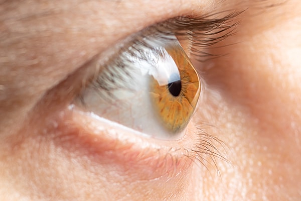
Cornea
Corneal Disease
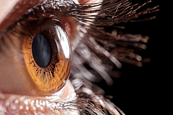
The cornea is a clear, thin dome-shaped tissue that composes the clear outer surface of the eye. It is the window in which light enters the eye. In a healthy eye, the cornea must have the correct shape and clarity to focus incoming light rays so they focus precisely onto the back of the eye. The cornea is comprised of five basic layers including the epithelium, Bowman’s membrane, the stroma, Descemet’s membrane and the endothelium.
Diseases and injuries that affect the cornea can cause pain and/or loss of vision. Certain conditions can cause the cornea to swell (called “edema”) and to become cloudy which reduces vision. Other conditions and injuries can distort or scar the cornea which causes visual distortions and reduced visual acuity. Symptoms and treatment can vary depending on the nature, location and severity of the condition.
Common diseases that affect the cornea can include:
Dry Eyes

Your eyes constantly produce tears so they stay moist and are comfortable. Some people do not produce enough tears or do not have the appropriate quality of tears to keep their eye comfortably moist. When this occurs, you may experience a variety of symptoms including:
- Dry and irritated eyes causing scratchiness
- Stinging
- Stringy mucus
- Eye fatigue and blurred vision
- Excess tearing
- Contact lens intolerance
- Sensitivity and irritation to smoke and wind
Dry eye is not only painful; it can be damaging to the eye’s tissues and impair vision. It is important for patients with this condition to take special care of their eyes in order to alleviate symptoms and prevent complications. Fortunately, many treatments options are available to help relieve symptoms and restore health back to the eyes.
Causes
Dry eye often increases with age as tear production slows, but the condition can also result from certain medications, conditions or injuries. Dry eye tends to affect women more often than men, as the hormonal changes that take place during pregnancy, menopause and with the effects of oral contraceptives can affect the consistency of tears. It is also more common in people over the age of 50. Other causes include:
- Medications such as antihistamines, decongestants, blood pressure medications and antidepressants
- Conditions such as Sjogren’s syndrome, rheumatoid arthritis, diabetes and thyroid problems
- Environmental conditions such as smoke, wind and dry climates
- Long-term contact lens use
- Refractive surgery
Treatments
Treatment for dry eye depends on the cause and severity of the condition, as well as the patient’s overall health and personal preference.
- Effective home remedies include blinking exercises, frequent breaks from close or computer work, avoiding wind and dry air from overheated rooms and blowing air, wearing sunglasses and increasing room humidity
- Artificial tears and ointments
- Prescription medications such as Restasis® & Xiidra™ to increase tear production and reduce inflammation
- Punctal plugs can be inserted into the corners of the eyes to limit tear drainage
- BlephEx® treatment for inflammatory dry eye
- Surgical closure of the drainage tubes of the eyes
- Eyelid surgery depending on the eyelid condition contributing to dry eyes
Keratoconus
Keratoconus is a progressive eye disease in which the dome-shaped cornea (the clear front window of the eye) becomes thin and develops a cone-like bulge (ectasia). As the condition progresses, the shape of the cornea is altered thus distorting your vision. Usually, keratoconus affects both eyes, although symptoms and progression in each eye may differ.
Keratoconus usually begins in the teenage years and early 20s, although it can develop at any age. Early symptoms include mild blurring of vision, increased sensitivity to light and glare, and mild eye irritation. As it progresses, the most common symptoms are increased blurring, increased nearsightedness or astigmatism, inability to wear contact lenses, and frequent eyeglass prescription changes.
Treatments
Early keratoconus is usually corrected with eyeglasses or soft contact lenses. However, as the condition progresses, treatment methods include specialty contact lenses, including scleral lenses, INTACS® (plastic implants that flatten the cornea), and Collagen Cross-linking (CXL). For more advanced keratoconus in which all other treatment methods have been exhausted, corneal transplant surgery can be performed.
Fuchs’ Corneal Dystrophy
Fuchs’ dystrophy is an inherited, progressive eye disease in which the cells in the cornea’s inner layer, called the endothelium, are reduced in number. This causes the remaining cells to swell or thicken. These cell changes may cause the cornea to become cloudy and swollen. Once the cells are lost, they do not grow back, so this condition will continue to progress with time. This condition affects both eyes and is more common in women than in men.
Symptoms and Treatments
Symptoms of Fuchs’ dystrophy include hazy or cloudy vision that develops in stages. In the early stage, as the cornea swells, vision in the morning may be hazy, but it clears up during the day. Treatment includes salt solution eye drops to help pull excess fluid from the cornea and reduce the swelling.
Once the disease has progressed to a more advanced stage, vision no longer clears, and instead, you may experience pain and sensitivity to light. Vision loss can begin to interfere with your life in the latter stages and prevents patients from functioning normally. In this stage, patients may require a partial thickness corneal transplant (DSAEK) to replace the diseased back layer of the cornea and allow clearer vision.
Pterygium
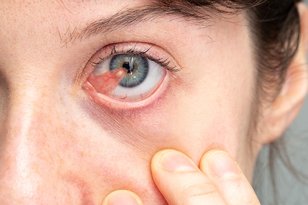
Pterygium is a benign fleshy growth of the conjunctiva (the lining of the white part of the eye) that grows over the cornea. It usually begins at the nasal cornea of the eye, but can appear in the outer corner as well. It may remain small or may grow large enough to interfere with vision. The exact cause of ptergia is not well understood, but it occurs more often in people who spend a lot of time outdoors, especially in sunny climates. Long-term sunlight exposure (UV rays), chronic eye irritation and dry eye play an important role.
Patients with pterygium often first notice the appearance of a lesion on their eye and can have dryness, itching, tearing or redness in that area. A pterygium that stays on the white part of the eye is termed a pingueculum. Once it begins to extend onto the cornea, it is termed a pterygium.
Prevention
Proper protection from UV rays is recommended, including sunglasses, especially ones that provide side coverage, and a large brimmed hat for outdoor activities. In hot, dry climates, artificial tears (eye drops) can be used to lubricate the eyes.
Treatment
In mild cases when a pterygium becomes red and irritated, artificial tears or ointments can be used to reduce inflammation. In more severe cases when the pterygium grows rapidly or is large enough to threaten sight, it can be removed surgically. Surgery is the only way to definitively remove a pterygium, but it is not a perfect solution. It requires long-term follow-up and despite proper surgical removal, it may return especially in young people.
- Conjunctival auto-grafting is a safe and effective technique to surgically remove a pterygium. It involves removing the pterygium tissue overlaying the sclera, and then replacing it with healthy conjunctival tissue obtained from the patient’s upper eyelid.
- Amniotic membrane transplantation is another safe and effective treatment in which the pterygium tissue is removed and amniotic placental tissue is used to reconstruct the surface of the eye. This type of graft encourages healing and reduces swelling.
Corneal Transplantation
There are several different corneal transplant procedures available to help to restore vision in patients with corneal problems. Traditional full thickness penetrating keratoplasty (PKP) involves removing all layers of the cornea and replacing the tissue with one from a healthy human donor, obtained from an eye bank. While this procedure is still performed, technological advances have allowed for the development of specialized corneal transplant procedures that replace only the damaged part of the cornea, while leaving the healthy parts intact. These procedures include:
- DALK: Deep anterior lamellar keratoplasty involves removing all anterior corneal tissue, while leaving the posterior corneal tissues (Descemet’s membrane and the endothelium) intact. This procedure is most useful in treating anterior corneal pathologies in which there is a normal functioning endothelium.
- DSAEK: Descemet’s stripping automated endothelial keratoplasty replaces the innermost layers of the cornea, known as the endothelium, while leaving the anterior corneal tissue intact. This procedure is performed through a much smaller incision with shorter recovery time and fewer risks than a traditional corneal transplant. DSAEK is commonly performed on patients with Fuchs’ Dystrophy.